

Óleo de peixe e óleos de sementes e grãos – algum motivo para consumi-los Ɂ
A "essencialidade" dos “óleos essenciais” de grãos e sementes [ômegas 6] e do óleo de peixe [ômega 3] - uma reflexão necessária.

[Imagem: zdravopharm.com.ua]
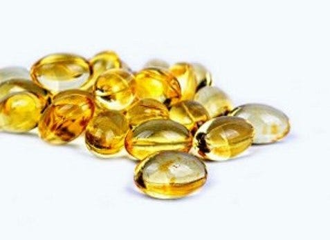
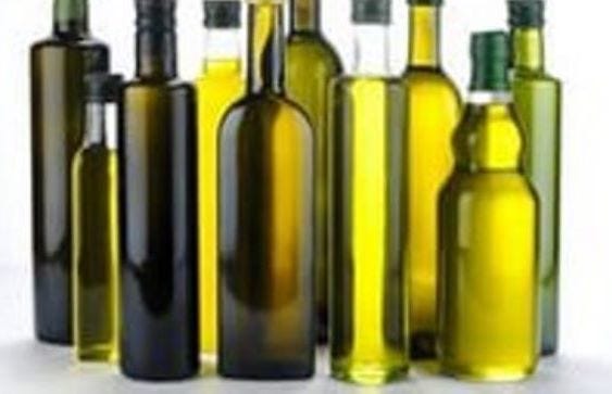
O óleo de peixe, assim como os óleos vegetais de sementes e grãos integram o cotidiano alimentar de meio mundo.
A corporação médica médico valida todos eles e existe uma indústria bilionária de alimentos e suplementos girando em torno desses óleos insaturados. Seu consumo, depois da II Guerra cresceu de forma exponencial.
Ele provavelmente estará na cozinha mais próxima e dez em dez restaurantes refogam, fritam ou temperam seus alimentos com tais óleos.
Mas o que muita gente não parou para observar é que esses óleos estragam muito facilmente. Sequer precisam ser fritos - como na confeção do pastel de feira - para se degradarem e se tornarem tóxicos.
Se a pessoa tiver o cuidado de deixar uma porção de quaisquer desses óleos na temperatura ambiente, do verão, por exemplo, definitivamente não vai gostar de ver o resultado. Todos eles se degradarão a olhos vistos: entram em processo de rancificação e até mudam de cheiro.
São facilmente oxidáveis fora da temperatura fria.
O óleo de peixe vem de animais que vivem em temperaturas muito frias, ligeiramente acima de zero, nas águas profundas. Como tais óleos [ômegas e no caso do peixe de águas profundas, o ômega 3] ficam fluidos a baixas temperaturas, resulta que são adequados para o metabolismo daqueles animais de sangue frio. Mas a simples lógica já sugere que não seriam convenientes para animais de sangue quente. E que seria de se esperar que eles se rancifiquem e sejam degradados no nosso corpo onde prevalecm temperaturas invariavelmente quentes.
Uma discussão aparentemente tão simples não costuma ser feita pela medicina oficial que continua propagando as virtudes do óleo de peixe e dos demais insaturados [ômega 6 e ômega 6] para a saúde humana.
Vejamos um breve comentário do pesquisador R. Peat [1936-2022] a respeito.
“Se você colocar um pedaço de gordura de peixe sobre a mesa, à temperatura ambiente, o efeito consegue ser ainda pior do que deixar óleo de canola ou de soja, milho igualmente expostos à temperatura ambiente. O óleo se torna rancificado muito rapidamente .
Esse fenômeno de o óleo de peixe estragar facilmente, tem a ver com o fato de que aqueles peixes evoluíram para viverem em água fria em uma temperatura ligeiramente acima do ponto de congelamento. Na temperatura ambiente, tais óleos começam a se oxidar rapidamente. Qualquer pessoa pode fazer uma experiência simples.
Se você dispuser de um litro de um óleo vegetal insaturado pode por uma cortiça em um tubo pequeno de borracha com o óleo e por em um copo de água. Na temperatura ambiente o óleo agirá como se estivesse respirando – ele estará queimando oxigênio e se degradando, se convertendo basicamente em um material tipo plástico ou verniz, e puxa a água para cima no tubo como se um pequeno organismo estivesse respirando na garrafa.
Óleo de peixe é ainda pior do que os óleos padrão de sementes. A não ser que seja um peixe do rio Amazonas onde a água é muito quente e aquele peixe possui gorduras que são mais seguras [semelhantes ao azeite de oliva], mas não encontramos tais peixes nas águas dos Estados Unidos [...] Todo organismo reflete a temperatura do seu tecido, com os tecidos quentes possuindo uma gordura mais saturada já que os óleos insaturados se degradam muito rapidamente em temperaturas mais quentes. Um bioquímico fez a experiência de colocar pulôvers em porcos e descobriu que sua gordura se tornava mais saturada, simplesmente por manter a pele aquecida”[R. Peat].
[A Wikipedia informa que rancificação significa a oxidação completa ou incompleta - ou a hidrólise - de gorduras quando expostas ao ar, luz” etc, mas náo cuida de informar que as gorduras que se rancificam facilmente são as insaturadas e, tampouco menciona o fator temperatura].
Certamente é possível que uma pessoa profundamente abduzida pela cultura pró-óleos de sementes, tipo soja, girassol, milho, linhaça, reaja ao comentário acima, de R Peat, alegando que se trata de uma anedota ou uma “mera” observação. E pode até acrescentar que “quem segue a ciência” sabe que o “mundo científico” aprova os óleos insaturados na alimentação humana há décadas e que são “essenciais” e por aí afora.
O problema sempre, no nosso tempo, é que “seguir a ciência” quando a ciência segue ou está atrelada ao cinheiro pode levar a conclusões problemáticas, verdadeiros pontos cegos.
E um deles, que tem o porte de uma verdadeira lenda, é o dos óleos insaturados.
Pesquisadores, há mais de 70 anos acumulam evidências contra o consumo de tais óleos, inclusive, naturalmente o óleo de peixe.
[Imagem: fancyface.ru]
Uma amostra bem minúscula de tais evidências pode ser conferida nas referências bibliográfica abaixo.
Mas como dizia um personagem do filme “A vida de Brian”, antes de mais nada é preciso dialogar com a realidade, com os fatos e não com o dogma.
Enquanto isso, enquanto esse debate não é ostensivamente aberto nas faculdades e entidades da área de saúde e nutrição, talvez caiba às pessoas que prezam por sua saúde, buscarem se informar por fora da lenda. Inclusive para constatarem se o pensamento de R. Peat sobre o tema é narrativa ou realidade.
GM Fontes, Goiânia, 27-6-23,
As informações aqui presentes não pretendem servir para uso diagnóstico, prescrição médica, tratamento, prevenção ou mitigação de qualquer doença humana. Não pretendem substituir a consulta ao profissional médico ou servir como recomendação para qualquer plano de tratamento. Trata-se de informações com fins estritamente educativos.
Referências________________
BRUDER E D BALL D L, 2003. Am J Physiol Regul Integr Comp Physiol. 2003 Jun;284(6):R1631-5. Epub 2003 Apr 10. An oxidized metabolite of linoleic acid stimulates rat ACTH (0, 0.2, or 2.0 ng/ml). In the absence of ACTH, EKODE (26 microM) increased corticosterone production from 5.3 +/- 2.3 to 14.7 +/- 5.0 ng. 10(6) cells. h(-1). The stimulatory effect of ACTH was increased threefold in the presence of EKODE (26.0 microM). Cholesterol transport/P-450scc activity was assessed by measuring basal and cAMP-stimulated pregnenolone production in the presence of cyanoketone (1.1 microM). EKODE (13.1 and 26.0 microM) significantly increased basal and cAMP-stimulated (0.1 mM) pregnenolone production. In contrast, EKODE decreased the effect of 1.0 mM cAMP. EKODE had no effect on early or late-pathway activity in isolated mitochondria. We conclude that EKODE stimulates corticosterone biosynthesis and amplifies the effect of ACTH. Increased levels of fatty acid metabolites may be involved in the increased glucocorticoid production observed in obese humans.
HILLERED, L, CHAN P H, 1989. J Neurosci Res . 1989 Oct;24(2):247-50. doi: 10.1002/jnr.490240216. Brain mitochondrial swelling induced by arachidonic acid and other long chain free fatty acids. L Hillered 1, P H Chan Polyunsaturated fatty acids (PUFAs), arachidonic acid in particular, are well known, potent inducers of edema in the brain, while monounsaturated and saturated long chain fatty acids do not possess this quality. This investigation has compared the ability of some free fatty acids (FFAs), known to be released during cerebral ischemia, to induce brain mitochondrial swelling in vitro. The PUFAs tested, especially arachidonic acid (20:4), were more potent in causing swelling than saturated or monounsaturated ones, as measured by the decrease in light absorbance of the mitochondrial suspension. This finding is in line with the unique potency of 20:4 to induce brain edema. Incubation of brain mitochondria with 20:4 for 20 min caused a dose-dependent swelling. [...] https://pubmed.ncbi.nlm.nih.gov/2531232/
HILLERED L, CHAN P H, 1998. Role of arachidonic acid and other free fatty acids in mitochondrial dysfunction in brain ischemia. Hillered L, Chan PH.J Neurosci Res. 1988 Aug;20(4):451-6. doi: 10.1002/jnr.490200407.PMID: 3141627
CHAN P H, FISHMAN R A, 1985. Cellular and molecular effects of polyunsaturated fatty acids in brain ischemia and injury. Chan PH, Fishman RA, Longar S, Chen S, Yu A.Prog Brain Res. 1985;63:227-35. doi: 10.1016/S0079-6123(08)61986-X.PMID: 3939039 Review. No abstract available.
PEAT, Ray, 2006. Suitable fats, unsuitable fats: issues in nutrition. In: raypeat.com/articles/
RUSYN I, 1999. Carcinogenesis. 1999 Nov;20(11):2095-100. Corn oil rapidly activates nuclear factor-kappaB in hepatic Kupffer cells by oxidant-dependent mechanisms. Rusyn I, Bradham CA, Cohn L, Schoonhoven R, Swenberg JA, Brenner DA, Thurman RG.
ANTHONY M, 1978. Res Clin Stud Headache 1978;6:110-6. Role of individual free fatty acids in migraine. Anthony M “Total plasma free fatty acids, platelet serotonin content and plasma stearic, palmitic, oleic and linoleic acids were estimated in 10 migraine patients before, during and after a migraine attack. Total and individual plasma free fatty acid levels rose and platelet serotonin content fell in most patients. The highest rise was observed in linoleic acid, which is known to be a potent liberator of platelet serotonin in vitro and is the only precursor of all prostaglandins in the body. It is suggested that the rise in plasma levels of linoleic acid in migraine could be responsible for the platelet serotonin release observed during the attack.” Citado por R. Peat.
GARCIA M C, KIM H Y, 1997. Brain Res 1997 Sep 12;768(1-2):43-8. Mobilization of arachidonate and docosahexaenoate by stimulation of the 5-HT2A receptor in rat C6 glioma cells. Garcia MC, Kim HY Laboratory of Membrane Biochemistry and Biophysics, National Institute on Alcohol Abuse and Alcoholism, National Institutes of Health, Rockville, MD 20852, USA. “In this study, we demonstrate that astroglial 5-HT2A receptors are linked to the mobilization of polyunsaturated fatty acids (PUFA). [...] “These results indicate that the 5-HT2A receptor is coupled to the mobilization of PUFA.” Citado por R. Peat.
KUJALOVA V, 1975. Bratisl Lek Listy 1975 Jul;64(1):58-63. [The effect of serotonin on the release of free fatty acids from human and rat adipose tissue (author's transl)]. [Article in Czech] Rath R, Kujalova V.
YOSHIDA, K ASAOKA Y, 1992. Proc Natl Acad Sci U S A 1992 Jul 15;89(14):6443-6. Platelet activation by simultaneous actions of diacylglycerol and unsaturated fatty acids. Yoshida K, Asaoka Y, Nishizuka Y “Several cis-unsaturated fatty acids such as oleic, linoleic, linolenic, eicosapentaenoic, and docosahexaenoic acids added directly to intact human platelets greatly enhance protein kinase C activation as judged by the phosphorylation of its specific endogenous substrate, a 47-kDa protein.” “In the presence of ionomycin and either 1,2-dioctanoylglycerol or phorbol 12-myristate 13-acetate, the release of serotonin from the platelets is also remarkably increased by cis-unsaturated fatty acids. The effect of these fatty acids is observed at concentrations less than 50 microM. Saturated fatty acids and trans-unsaturated fatty acids are inactive.” “. . . cis-unsaturated fatty acids increase an apparent sensitivity of the platelet response to Ca2+. The results suggest that cis-unsaturated fatty acids, which are presumably produced from phosphatidylcholine by signal-dependent activation of phospholipase A2, may take part directly in cell signaling through the protein kinase C pathway.” Citado por R. Peat.
TAPPIA W J, 1995. Influence of unsaturated fatty acids on the production of tumour necrosis factor and interleukin-6 by rat peritoneal macrophages, Mol. Cell Biochem. 143(2), 89-98, 1995.
VANPAPENDORP D.H. et al. 1995. , Biochemical profile of osteoporotic patients on essential fatty supplementation, Nutr. Res. 15(3), 325-334, 1995. (Fish oil increased urinary calcium fish oil/evening primrose oil increased osteocalcin and procollagen.)
FELTON C V, et al., 1994. Dietary polyunsaturated fatty acids and composition of human aortic plaques, Lancet 344(8931), 1195-1196, 1994.
DARMIANI H, et al., 1994. Interferon-gamma and polyunsaturated fatty acids increase the binding of lipopolysaccharide to macrophages, Int. J. Exp. Pathol. 75(5), 363-368, 1994.
RAFAEL, J, et al., 1984. The effect of essential fatty acid deficiency on basal respiration and function of liver mitochondria in rats, J. Nutr. 114, 255-262, 1984.
CHAN P.H. FISHMAN, R A, 1978. Brain edema: Induction in cortical slices by polyunsaturated fatty acids, Science 201, 358-369, 1978. “Ibis cellular edema was specific, since neither saturated fatty acids nor a fatty acid containing a single double bond had such effect.” Citado por R. Peat.
LARSSON B, et al., 1995. Effects of dietary alpha- and gamma-linolenic acids on liver fatty acids, lipid metabolism, and survival in sepsis, Shock 4(l), 11-20, 1995. “Dietary GLA reduced survival from sepsis.”
CHEMLA D, et al., 1995. Influence of dietary polyunsaturated fatty acids on contractility, inotropy and compliance of isolated rat myocardium, J mol Cell Cardiol 27(8), 1745-1755, 1995. “There was a trend towards a lower peak lengthening velocity at preload in the LC (n-3) group … together with an unchanged peak rate of isometric force decline. This resulted in a significant impairment of the two mechanical indexes testing the load dependence of myocardial relaxation.” See B. Pieske, Circul. 92(5),1169-78. Citado por R. Peat.
ENDRESEN M.J. et al., 1994. Effects of free fatty acids found increased in women who develop pre-eclampsia on the ability of endothelial cells to produce prostacyclin, oGMP and inhibit platelet aggregation, Scan. J. Clin. Lab. Invest. 54(7), 549-557, 1994. “…levels of circulating fire fatty acids are increased in women who later develop pre-eclampsia long before the clinical onset of the disease.” “…linoleic acid reduced the thrombin-stimulated prostacyclin release by 30-60% oleic acid by 10-30%, wheras palmitic acid had no effect.” “Linoleic acid reduced the endothelial cells’ ability to inhibit platelet aggregation by 10-45%….” Citado por R. Peat.
D´AQUINO M. et al., 1991. Effect offish oil and coconut oil on antioxidant defence system and lipid peroxidation in rat liver, Free Radical Res. Commun. (Switzerland) 12-13 (1), 147-152, 1991. The rate of lipid peroxidation in isolated microsomes was three-fold higher in rats fed fish oil as compared to rats with coconut oil diet.” “These results suggest that fish oil feeding at an amount compatible with human diet, although decreasing plasma lipids, actually challenges the antioxidant defence system, thus increasing the susceptibility of tissues to free radical oxidative damage.” Citado por R. Peat
WANG Y.P. 1995. Aspirin inhibits both lipid peroxides and thromboxane in preeclamptic placentas, Free Radical Biol. Med. 18(3), 585-591, 1995.
BANGUR C.S. 1995. Thyroid hormone treatment alters phospholipid composition and membrane fluidity of rat brain mitochondria, Biochem. J. 305(1),29-32, 1995. (Increases fluidity.)
SOHAL et al., 1995. Mitochondrial superoxide and hydrogen peroxide generation, protein oxidative damage,and longevity in different species of flies, Free Rad. Biol. & Med. 19(4),499-504, 1995. Cytochrome C oxidase protects against free radical damage. This enzyme depends on thyroid and light. Citado por R. Peat
ODA E HATADA K, 2005. Int Heart J. 2005 Nov;46(6):975-85. Relationships between serum unsaturated fatty acids and coronary risk factors: negative relations between nervonic acid and obesity-related risk factors. Oda E, Hatada K, Kimura J, Aizawa Y, Thanikachalam PV, Watanabe K. "The objective of the present study was to analyze the relationships between serum USFA and CRF [coronary risk factors]." "Oleic acid (OA), linoleic acid (LA), and eicosapentaenoic acid (EPA) were positively related to coronary risk factors (total CRFS = 2, 3, and 4, respectively), while nervonic acid (NA) exerted negative effects on these risk factors (total CRFS = -6 ). It is concluded NA may have preventive effects on obesity-related metabolic disorders."
OMURA M, 2001. FEBS Lett. 2001 Jan 5;487(3):361-6. Eicosapentaenoic acid (EPA) induces Ca(2+)-independent activation and translocation of endothelial nitric oxide synthase and endothelium-dependent vasorelaxation. Omura M, Kobayashi S, Mizukami Y, Mogami K, Todoroki-Ikeda N, Miyake T, Matsuzaki M. "EPA stimulated NO production even in endothelial cells in situ loaded with a cytosolic Ca(2+) chelator . . . which abolished the [Ca(2+)]i elevations induced by ATP and EPA."
TSOUTSIKOS P MINERS J O, 2004. Biochem Pharmacol. 2004 Jan 1;67(1):191-9. Evidence that unsaturated fatty acids are potent inhibitors of renal UDP-glucuronosyltransferases (UGT): kinetic studies using human kidney cortical microsomes and recombinant UGT1A9 and UGT2B7. Tsoutsikos P, Miners JO, Stapleton A, Thomas A, Sallustio BC, Knights KM.
AYRE K J HULBERR A J, 1997. Lipids. 1997 Dec;32(12):1265-70. Dietary fatty acid profile affects endurance in rats. Ayre KJ, Hulbert AJ. "The diets comprised an essential fatty acid-deficient diet (containing mainly saturated fatty acids); a diet high in n-6 fatty acids, High n-6; and a diet enriched with n-3 fatty acids, High n-3. Submaximal endurance in rats fed the High n-3 diet was 44% less than in rats fed the High n-6 diet (P < 0.02). All rats were then fed a standard commercial laboratory diet for a 6-wk recovery period, and their performances were reevaluated. Although endurance in all groups was lower then at 9 wk, it was again significantly 50% lower in the High n-3 group than the High n-6 group (P < 0.005). Although n-3 fats are considered beneficial for cardiovascular health, they appear to reduce endurance times, and their side effects need to be further investigated." Citado por R. Peat.
BARTFAI E, ORSIERE T, 2000. Ann Biol Clin (Paris) 2000 Sep-Oct;58(5):595-600. [Studies on the genotoxic effects of crude liver oils from 3 species of Mediterranean sharks by means of in vitro micronucleus test using human lymphocytes] Bartfai E, Orsiere T, Duffaud F, Villani P, Pompili J, Botta A. "The results of this experimental study show that the crude liver oils of three species of sharks are genotoxic and confirm a high carcinogenic risk." Citado por R. Peat.
DHEIN S MICHAELIS B, 2005. Naunyn Schmiedebergs Arch Pharmacol. 2005 Mar;371(3):202-11. Epub 2005 Apr 15. Antiarrhythmic and electrophysiological effects of long-chain omega-3 polyunsaturated fatty acids. Dhein S, Michaelis B, Mohr FW. “Atrioventricular conduction time was slowed only by DHA and EPA.” “Regarding antiarrhythmic activity we found that the threshold for elicitation of a ventricular extrasystole was concentration-dependently enhanced by DHA and EPA, but not by ALA. DHA dose-dependently reduced longitudinal propagation velocity V(L) and to a lower extent transverse velocity V(T).”
DIAZ O BERQUAND A, 2002. J Biol Chem. 2002 Oct 18;277(42):39368-78. The mechanism of docosahexaenoic acid-induced phospholipase D activation inhuman lymphocytes involves exclusion of the enzyme from lipid rafts. Diaz O, Berquand A, Dubois M, Di Agostino S, Sette C, Bourgoin S, Lagarde M, Nemoz G, Prigent AF. “Docosahexaenoic acid (DHA), an n-3 polyunsaturated fatty acid that inhibits T lymphocyte activation, has been shown to stimulate phospholipase D (PLD) activity in stimulated human peripheral blood mononuclear cells (PBMC).” “This PLD activation might be responsible for the immunosuppressive effect of DHA because it is known to transmit antiproliferative signals in lymphoid cells.” Citado por R. Peat
GAIVA M H, COUTO R C, 2003. Nutrition. 2003 Feb;19(2):144-9. Diets rich in polyunsaturated fatty acids: effect on hepatic metabolism in rats. Gaiva MH, Couto RC, Oyama LM, Couto GE, Silveira VL, Ribeiro EB, Nascimento CM. “Male Wistar rats, just weaned, were fed ad libitum for 8 wk with one of the following diets: rat chow (C), rat chow containing 15% (w/w) soybean oil (S), rat chow containing 15% (w/w) fish oil (F), and rat chow containing 15% soy bean and fish oil (SF; 5:1, w/w).” “Body weight gain was higher in F and SF than in C and S rats. Liver weight, lipid content, and lipogenesis rate increased in F and SF rats, although adenosine triphosphate citrate lyase activity decreased. Glycogen concentration decreased in S, F, and SF rats compared with C rats.” Citado por R. Peat.
GAIVA M H COUTO R C, 2001. Br J Nutr. 2001 Sep;86(3):371-7. Polyunsaturated fatty acid-rich diets: effect on adipose tissue metabolism in rats. Gaiva MH, Couto RC, Oyama LM, Couto GE, Silveira VL, Riberio EB, Nascimento CM. “Wistar rats were fed ad libitum, for 8 weeks with one of the following diets: C, rat chow; S, rat chow containing 15 % (w/w) soyabean oil; F, rat chow containing 15 % (w/w) fish oil; SF, rat chow containing 15 % (w/w) soyabean and fish oil (5:1, w/w).” "Energy intake was reduced while carcass lipid content was increased in the three fat-fed groups." "These results indicate that enrichment of the diet with polyunsaturated fatty acids causes changes in adipose tissue metabolism that favour fat deposition. Different metabolic pathways were preferentially affected by each type of fatty acid used."
HOUSSAY B A, 1947. B. A. Houssay and C. Martinez, Experimental diabetes and diet, Science 105, 548-549, 1947.
HULBERT A J, 2005. J Theor Biol. 2005 May 21;234(2):277-88. On the importance of fatty acid composition of membranes for aging. Hulbert AJ. Mech Ageing Dev. 2006 Apr 16; Extended longevity of wild-derived mice is associated with peroxidation-resistant membranes. Hulbert AJ, Faulks SC, Harper JM, Miller RA, Buffenstein R. "Muscle and liver phospholipids from these long-living mice lines have a reduced amount of the highly polyunsaturated omega-3 docosahexaenoic acid compared to the DC mice, and consequently their membranes are less likely to peroxidative damage. The relationship between maximum longevity and membrane peroxidation index is similar for these mice lines as previously observed for mammals in general. It is suggested that peroxidation-resistant membranes may be an important component of extended longevity."
KAJIHARA H TOTOVIC V, 1975. Virchows Arch B Cell Pathol. 1975 Nov 21;19(3):239-54. [Ultrastructure and morphogenesis of ceroid pigment. II. Late changes of lysosomes in Kupffer cells of rat liver after phagocytosis of unsaturated lipids] Kajihara H, Totovic V, Gedigk P. "These lipids, which have been changed in their molecular structure, cannot be hydrolized by lysosomal enzymes. They remain as an indigestible material, as a waste product in lysosomal residual bodies. Both lipofuscin and ceroid are lysosomal structures containing oxidized and polymerized lipids."
MARTINEZ M BALLABRIGA A, 1987. Lipids 22(3), 133-6, 1987. Effects of parenteral nutrition with high doses of linoleate on the developing human liver and brain, Martinez, M., and A. Ballabriga.
FEBS Lett. 1998 Oct 16;437(1-2):24-8. Generation of protein carbonyls by glycoxidation and lipoxidation reactions with autoxidation products of ascorbic acid and polyunsaturated fatty acids. Miyata T, Inagi R, Asahi K, Yamada Y, Horie K, Sakai H, Uchida K, Kurokawa K. Citado por R. Peat.
REITHMANN C, 1996. Naunyn Schmiedebergs Arch Pharmacol. 1996 Jul;354(2):109-19. Exposure to the n-3 polyunsaturated fatty acid docosahexaenoic acid impairs alpha 1-adrenoceptor-mediated contractile responses and inositol phosphate formation in rat cardiomyocytes. Reithmann C, Scheininger C, Bulgan T, Werdan K. “The results presented show that chronic n-3 polyunsaturated fatty acid pretreatment of rat cardiomyocytes leads to a marked impairment of alpha 1-adrenoceptor-induced positive inotropic effects and induction of arrhythmias concomitant with a n-3 fatty acid-induced decrease in IP3 formation.” Citado por R. Peat
SAKAMORO, N NISHIIKE, T, 2000. Nutrition. 2000 Jan;16(1):11-4. Effects of eicosapentaenoic acid intake on plasma fibrinolytic and coagulation activity by using physical load in the young. Sakamoto N, Nishiike T, Iguchi H, Sakamoto K. “Thus, as determined by the load, a small amount of daily EPA intake clearly decreased fibrinolytic activity and increased coagulation activity.” Citado por R. Peat
Nutr Cancer 1998;30(2):137-43. Effects of dietary n-3-to-n-6 polyunsaturated fatty acid ratio on mammary carcinogenesis in rats. Sasaki T, Kobayashi Y, Shimizu J, Wada M, In'nami S, Kanke Y, Takita T. "Dietary fat was fed to the rats as 10% of the total feed weight, starting two weeks before the initiation. An increase in the n-3/n-6 ratio did not suppress the incidence or reduce the latency of mammary tumor development. The number and weight of mammary tumors per tumor-bearing rat tended to be large in the group with an n-3/n-6 ratio of 7.84 compared with those in the other groups. As the n-3/n-6 ratios were elevated, the total number and weight of tumors increased gradually." "These results suggested that the increase in the n-3/n-6 ratio of dietary fat with the fixed PUFA-to-saturated fatty acid ratio cannot suppress the mammary carcinogenesis but can promote development of tumors, despite reduced PGE2 concentration in the tumor." Citado por R. Peat.
SONG J H FUJIMOTO K, 2000. J Nutr 2000 Dec;130(12):3028-33. Polyunsaturated (n-3) fatty acids susceptible to peroxidation are increased in plasma and tissue lipids of rats fed docosahexaenoic acid-containing oils. Song JH, Fujimoto K, Miyazawa T.. "Thus, high incorporation of (n-3) fatty acids (mainly DHA) into plasma and tissue lipids due to DHA-containing oil ingestion may undesirably affect tissues by enhancing susceptibility of membranes to lipid peroxidation and by disrupting the antioxidant system."
STEERENBERG, P A, 2002. Diabetes Nutr Metab. 2002 Aug;15(4):205-14. Long-term effect of fish oil diet on basal and stimulated plasma glucose and insulin levels in ob/ob mice. Steerenberg PA, Beekhof PK, Feskens EJ, Lips CJ, Hoppener JW, Beems RB. “We have investigated, in comparison to low and high fat diets, the effect of a fish oil diet on basal and stimulated plasma glucose and insulin levels in male and female ob/ob mice.” “Intercurrent deaths were found especially in the fish oil diet group. Compared to the other diet groups, plasma insulin levels of the fish oil diet group were significantly increased 3 months after the start of the diet and remained higher for another 3 months.” “At 12 months, microscopy revealed an increased severity of hepatic brown pigment accumulation and extramedullary haematopoiesis in the spleen of mice fed with fish oil.” “Fish oil diet also increased intercurrent mortality. However, a consistent course of death could not be established using morphological parameters.” Citado por R. Peat
[A74] J Biol Chem. 2002 Feb 15;277(7):5692-7. Unsaturated fatty acids inhibit cholesterol efflux from macrophages by increasing degradation of ATP-binding cassette transporter A1. Wang Y, Oram JF. “These findings raise the possibility that an increased supply of unsaturated fatty acids in the artery wall promotes atherogenesis by impairing the ABCA1 cholesterol secretory pathway in macrophages.” Citado por R. Peat
***